The term “genodermatoses” applies to skin conditions of genetic origin. Genodermatoses are most often congenital (present at birth), but appearance may be delayed. Additionally, Genodermatoses may be hereditary or due to some influence during gestation, even up to the moment of birth.
Dermatomyositis
Familial canine dermatomyositis is a hereditary inflammatory condition of the skin and muscle of young Collies, Shetland sheepdogs, Beauceron shepherds, and their crosses. It has also been reported in the Welsh Corgi, Lakeland Terrier, Chow Chow, German Shepherd Dog, and Kuvasz. The familial basis in these other breeds is not proven. Breeding studies in Collies and Shetland sheepdog support an autosomal dominant mode of inheritance. The cause is unknown. A genetically determined immune-mediated pathogenesis is suspected.
Lesions occur early in life, often before 6 months of age. Signs may be observed as early as 7-11 weeks of age. Progression of lesions is variable, but the extent of disease development is often known by 1 year of age. Lesions usually decrease in number, extent, and severity thereafter. There is no sex, coat color, or coat link association.
Lesions develop in areas of friction or mechanical trauma. Affected body sites include the face (especially around the eyes and mouth), ear tips, tail, carpal and tarsal regions of the limbs, digits, nails, and footpads. Lesions include: alopecia, erythema, scale, crusting, and ulceration. Vesicles may be present.
Muscle involvement occurs after skin lesions and correlates with the severity of skin lesions. The owner may observe dirty water bowls with food particles since the dogs don’t swallow completely. They may have difficulty drinking or prehending food. A high stepping or stiff gait may be present. Megaesophagus (large dilated esophagus) with resulting aspiration pneumonia may occur. The most common sign of the myositis is asymptomatic atrophy of the muscles of mastication (on the head) and distal limbs.
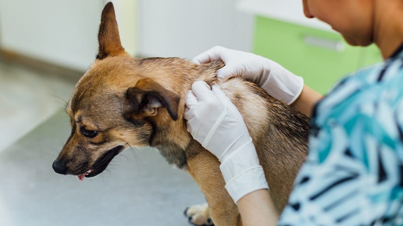
The diagnosis is made by ruling out other dermatoses with similar features in addition to the signalment and history. Biopsy is used to confirm the clinical diagnosis. Muscle biopsy and muscle tests may be performed.
Recommendations for treatment would include limiting mechanical trauma and sun exposure. Mild skin lesions heal spontaneously without intervention. Permanent scarring and alopecia may develop. Moderately affected dogs may be maintained as pets for prolonged periods with careful management. Treatments that have been prescribed include Vitamin E, Fatty acid supplementation, corticosteroids, and pentoxiphylline. The recommendation for humane euthanasia may be made for severely affected dogs, as they have widespread skin lesions, difficulty walking, eating, and drinking, and frequently develop aspiration pneumonia.
Sebaceous Adenitis
Sebaceous adenitis (SA) is an uncommon dermatosis recognized in many breeds that results in the destruction of the sebaceous glands. Sebaceous glands are part of the hair follicle unit and are important in the maintenance of the hair coat. SA is most commonly associated with the Standard Poodle, but the Gordon setter, Chow Chow, Akita, Golden Retriever, Viszla, Shih-tzu, and Havanese are others we often see with sebaceous adenitis.
In the standard poodle, pedigree analysis suggests that SA is a genetic disease with autosomal recessive mode of inheritance. Causal theories include: 1) sebaceous gland destruction is a developmental and inherited defect; 2) sebaceous gland destruction is due to cell-mediated immunologic reaction directed against a component of the gland; 3) the initial defect is a keratinization abnormality with subsequent obstruction of the sebaceous ducts resulting in inflammation of the glands; and 4) the sebaceous adenitis and keratinization defects are the result of an abnormality in lipid metabolism.
Breeding subclinically affected dogs offers the same risk of producing affected offspring as breeding dogs with observable disease. This indicates that subclinical animals are not carriers, but have the disease. It has been reported that biopsies from clinically normal standard Poodles that were still normal two years later may contain focal areas of SA.
There is no sex or coat color predilection, and the disorder tends to appear in young adult to middle aged dogs. 90% of lesions are recognized between 1.5 and 5 years of age. Over 25% of dogs diagnosed may have a subclinical form.
Typical presentation is dry scaling skin with patchy hair loss involving the ear flaps, dorsal trunk, dorsal neck, and top of the head. With time generalized disease is common. Scales may be tightly adhered to the hair shaft, and the coat may appear dull and brittle with or without a brown-red tint. Ear infections may occur. Secondary bacterial infections may be present. Itch is variable, but usually not pronounced. Severity is variable.
Signalment, history, and clinical signs form the basis of diagnosis. The veterinarian begins the diagnostic phase by ruling out more common skin diseases with similar clinical signs. Dermatohistopathology, biopsy of affected skin, confirms the diagnosis in affected dogs.
The principle of topical therapy is aimed at externally replacing the actions of the sebaceous gland with treatment. Topical therapy is relatively safe and may reduce scaling, restore coat luster, and reduce hair loss. Treatment of secondary infections alleviates patient discomfort if present. Systemic therapy may be recommended and modified cyclosporine (Atopica®, Novartis) is the medication typically prescribed. The findings on the skin biopsy would dictate whether modified cyclosporine is appropriate in the individual patient. Therapy is lifelong.
Atopic Dermatitis
Atopic dermatitis (AD) is a genetically-predisposed inflammatory and pruritic allergic skin disease with characteristic clinical features associated most commonly with IgE antibodies to environmental allergens.
It is generally felt that canine AD has a genetic component due to the clinical observation by veterinarians and veterinary dermatologists that the disease appears more often within certain breeds. However, one must consider the incidence of disease to the clinic population of those breeds (i.e. regional breed popularity.
AD is an allergic disease in which individuals develop hypersensitivity to pollens (trees, grasses, weeds), epidermals (cat, horse, sheep, cow, and human dander), fibers (cotton and feather), house dust mites, storage (grains) mites, and molds. Sensitization to allergens occurs through percutaneous absorption of allergen through the skin. Disease manifestation is the result of the hypersensitivity to these allergens.
Breeds that have been reported to have a high prevalence of AD include the West Highland White Terrier, Bichon Friese, Scottish Terrier, Dalmatian, Chinese Shar Pei, Boxers, Labrador and Golden Retriever, English Setters, German Shepherd Dogs, and Cocker Spaniels. Others have been reported, and prevalence within a breed likely depends on the breed popularity in the area from which the base population being evaluated is taken.