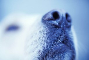
Mucocutaneous pyoderma (MCP) is a common condition that may affect the nasal planum in dogs. MCP is a bacterial infection usually caused by Staphylococcus pseudintermedius. German shepherd dogs are the most commonly affected breed.
MCP primarily affects the mucocutaneous junctions of the nasal planum and lips, but other mucocutaneous junction sites may be involved. If the nasal planum is affected, there is initial erythema and swelling of the nasal planum, typically at the alar folds, followed by crusts, then fissures and erosions. Beneath elevated crusts there is thick, purulent exudate and an erosive to ulcerative dermatitis.
Diagnosis of Mucocutaneous Pyoderma (MCP)
Diagnosis is made via dermatologic examination, cytology and response to treatment. Cytology of exudate reveals bacteria and inflammatory cells. Bacterial culture and susceptibility testing of purulent exudate from under a protected crust is indicated if there is a failed response to appropriate antibiotic therapy. If cytology does not reveal bacteria, a bacterial culture is sterile and there is a failed response to antimicrobial therapy, submission of a tissue biopsy for dermatohistopathology is indicated, as discoid lupus erythematosus is a condition with similar clinical appearance. A fungal culture may also be indicated, as some fungal diseases share clinical features with MCP.
Treatment of Mucocutaneous Pyoderma (MCP)
Treatment of MCP involves topical therapies and systemic antibiotics. Maintenance topical therapy is recommended for long-term control. Trichophyton dermatophytosis is one of the most common fungal infections we see affecting the nasal planum in dogs. This condition is caused by infection with the soil-borne dermatophyte Trichophyton mentagrophytes and dogs that hunt or root in soil, such as the Jack Russell terrier, are at an increased risk of developing the disease. Lesions of the skin and nasal planum may include, alopecia, erythema, papules, scaling, crusting, erosions or ulcerations. The clinical presentation may resemble autoimmune diseases such as discoid lupus erythematosus, pemphigus erythematosus or dermatomyositis. Pruritus of lesional sites varies from absent to severe. Diagnosis is via dermatologic examination, skin scrapings, cytology, culture and dermatohistopathology.