Calcinosis cutis (CC) is the deposition of inorganic, insoluble mineral salts into the different layers of the skin. It is characterized histologically (under the microscope in tissue samples) by mineralization of dermal collagen fibers, accumulation of macrophages and mixed inflammatory infiltrates, acanthosis (thickening of skin), ulceration and evidence of elimination of mineral through the skin.
Treatment for Calcinosis Cutis in Dogs
Calcinosis cutis is classified based upon the cause. Metastatic CC occurs when calcium salts precipitate within normal uninjured tissues. Hypercalcemia and hyperphosphatemia are the cause and are the result of underlying systemic calcium or phosphorous imbalance/metabolism in which the extracellular calcium overwhelms the cells ability to regulate intracellular calcium. It occurs with impaired kidney function and other inflammatory conditions that impair calcium metabolism.
Iatrogenic CC occurs from the injection of calcium containing products into the skin (IV administration of calcium chloride and gluconate) with material getting outside the vessel, skin absorption of calcium containing landscape products, or some medications. Idiopathic CC has no underlying cause and will resolve on its own within 12 months. Dystrophic CC is the most common type and caused by iatrogenic hyperadrenocorticism or spontaneous hyperadrenocorticism (pituitary or adrenal dependent). It also may occur with inflammatory skin conditions or diabetes mellitus. How dystrophic CC occurs is unclear, but gluconeogenic and protein catabolic activity in the skin involve cortisol. Cortisol is the body’s own steroid hormone produced in excess in these conditions. For dystrophic CC to occur there must be cation and anion deposition in to tissues and changes in collagen. Collagen changes occur from tissue injury and damage to cell membranes and result in transformation of minerals from dissolved ions in to solid phase. This causes an influx of calcium into cells that lowers the extracellular pH and impairs mineralization inhibitors. With more calcium flowing in to the cells, there is a rearrangement of the molecular structure of proteins and a formation of an organic matrix that attracts and binds the calcium. The result is mineralization of skin.
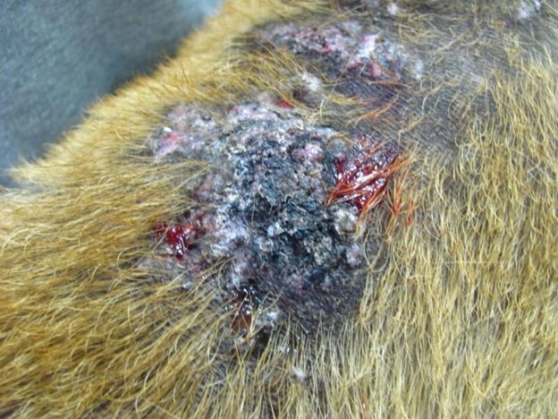
Lesions in CC are characterized by erythematous papules, plaques and nodules that are typically ulcerated and secondarily infected. Lesions are often found on the top of the trunk and head, as well as the axillae, ventrum, elbows, hocks, and oral cavity. Soft tissue organs such as the spleen and kidneys may also show mineralization. Calcinosis cutis usually resolves with removal of the underlying cause. It is important to treat all secondary infections. Frequent bathing in medicated shampoos and hydrotherapy are helpful. Sometimes, surgical removal of isolated lesions may be recommended if the surgeon feels wound healing would be successful in the individual patient. Recently, the use of minocycline for treatment of CC was reported. Though minocycline is an antibiotic, it chelates calcium and directly inhibits collagenolytic enzymes. Treatment with DMSO gel may hasten resolution of lesions. A thin film is applied one to two times a day. If glucocorticoids are being used and are the cause, they must be tapered and discontinued. Resolution is not immediate and takes time. The skin often looks worse before it looks better.
Image Credit: Vetbook.org