Pemphigus foliaceus (PF) is the most common feline autoimmune skin disease. Pemphigus diseases result from the formation of antibodies against keratinocyte structures, and differ because each has a different target antigen and/or histopathologic feature.
Epidermal cells have structures involved in cell-to-cell adhesion (desmosomes) or in cell-matrix adhesion (hemidesmosomes-anchoring fibrils complex). The desmosome is the component that holds keratinocytes (skin cells) together and is one structure in a dynamic system that support and connect cell to cell. They essentially link the cytoskeleton of one keratinocyte with that of its neighbor. Many proteins make up the desmosome.
When autoantibodies target a desmosome protein, a pemphigus disease results. The target protein in feline PF has not been identified. Binding of autoantibody to the target protein between keratinocytes during assembly and disassembly of the desmosome activates the keratinocyte. As a result, there is disruption of intercellular adhesion. Because keratinocytes lose the ability to remain attached to each other, they separate, lift off and detach forming blisters/pustules (acantholysis).
Production of autoantibodies in PF may be due to abnormal immune regulation or abnormal antigenic stimulation. Genetic factors may be involved, as cases have been described in littermates. Ultraviolet light may be a trigger factor. The autoimmune response may be caused, in some cases, by drug reactions. Drugs speculated to cause PF have included itraconazole, lime sulfur dip, ipodate, and methimazole, amoxicillin, and sulphonamides. A Siamese cat given cimetidine developed an exfoliative dermatitis of the ears, face, and feet. Footpad lesions and paronychia were also present. PF was the histopathologic diagnosis. The strength of evidence supporting drug causation remains weak.
History and Signalment
PF may affect cats of any age depending on the cause; median age 5 years. Domestic short hair cats are most often affected. Other reported breeds include domestic long-haired cats, Siamese, Himalayan, Persian, Maine Coon, American Blue, Somali, Scottish Fold, and Ragamuffin. The duration of disease prior to presentation varies. Previous dermatologic disease unassociated with the PF may be reported. It has been suggested that chronic inflammatory skin disease may play a causative role.
Lesions and Clinical Signs
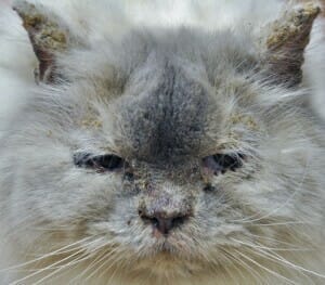
Feline PF varies in the severity and extent of lesions. The phenotype is usually mild and localized, but generalized lesions can occur. The primary lesion of PF is a pustule. Visibly intact pustules are rarely observed, as these superficial pustules are fragile and rupture easily forming crust. Crusting dermatitis is the common lesion. There is a tendency for lesions to occur in a bilaterally symmetrical pattern. Affected sites may include the face, neck, bridge of the nose, periocular skin, pinnae, paw pads, claw folds, periareolar skin, perivulvar skin, periscrotal skin, perineal skin, chin, dorsum, ventrum. Paronychia with a concurrent purulent exudate exuding from claw folds is a common clinical finding in cats, and periareolar crusting is frequently recognized as a characteristic predilection site. Pruritus is often reported, and otitis externa is common. Lethargy, anorexia, weight loss, lymphadenopathy, and elevated rectal temperature are often identified.
Diagnosis
Clinical signs, characteristic acantholytic cells identified from lesion cytology, elimination of other pustular to crusting skin diseases from the differential list, and compatible dermatohistopathology are used to confirm one’s clinical suspicion of feline PF. The hallmark of PF is the presence of acantholysis in dermatohistopathology samples. Diagnostic tests that should be performed include cytology, surface skin scrapings, and deep skin scrapings. If no problem is identified with these screening tests, and there is a clinical suspicion for feline PF, biopsies should be collected for dermatohistopathology and cultures should be submitted. Cultures are recommended to “prove” a sterile disease process. General laboratory tests are recommended but are most useful in assessing the overall health status of the cat as you plan for long term treatment with immunosuppressive medications.
Cytology
Demonstration of acantholytic cells from careful collection of pus from an intact pustule or from an impression of the eroded skin under an elevated crust increases the clinical suspicion for PF. One will often see clustered rafts of acantholytic cells or free-floating ones admixed with neutrophils and rarer eosinophils. The presence of acantholytic cells is not specific for PF, however. Dermatophytosis and bacterial folliculitis can result in acantholysis.
Dermatohistopathology
Histopathologic evaluation of biopsied lesions has the potential to give the most valuable diagnostic information. Pustules are the ideal lesion for submission, but a crusted lesion is the second-best choice. The dermatohistopathologist will provide a description that will identify typical patterns such as: Pustular dermatitis, hyperplastic dermatitis, orthokeratotic hyperkeratosis with focal parakeratosis, perivascular to interstitial neutrophilic and eosinophilic dermatitis, and perhaps apoptosis of keratinocytes.
Treatment of Feline PF
Treatment requires immune suppression and immunomodulation. Do not treat pemphigus foliaceus without a diagnosis and elimination of other differentials.
Glucocorticoids are the most common initial treatment, as they are potent anti-inflammatory and immunomodulatory agents. There are numerous mechanisms of action that are useful in the treatment of autoimmune skin diseases, but the most notable are the effect on humoral and cell-mediated immunity, phagocyte defenses, inhibition of inflammatory mediators, and suppression of autoantibody levels.
In the author’s experience in treatment of feline PF, glucocorticoid monotherapy has been effective in achieving clinical remission in most cases. Prednisone, prednisolone, triamcinolone, methylprednisolone, and dexamethasone are most widely used. Prednisolone and not prednisone should be used in cats based upon the assessment of some recent pharmacologic data. It has been shown that oral prednisone is not well absorbed and/or converted to the active form of prednisolone in cats. Some authors prefer triamcinolone or dexamethasone to prednisone. The glucocorticoid chosen should be administered at immunosuppressive dosages until remission is achieved (2-8 weeks) and tapered to the lowest effective alternate day dosage for maintenance therapy. If side effects are undesirable, or if there is no significant improvement upon reevaluation at 2 and 4 weeks, an alternate glucocorticoid should be selected, or a nonsteroidal immunosuppressant should be added. I do not provide dosages here because standard references provide accepted dosing ranges.
Chlorambucil is most often used in cats when glucocorticoid monotherapy is not sufficient. One must monitor for myelosuppression in addition to vomiting, diarrhea, and anorexia. CBC with platelet count recommended before treatment and as indicated throughout treatment.
Chyrsotherapy is the use of gold salts. The use of chrysotherapy and glucocorticoids has been shown to be effective in the treatment of feline PF. Gold salts have immunomodulating and anti-inflammatory effects.
Cyclosporine is a nonsteroidal immunosuppressive drug that is sometimes prescribed. The clinical response is variable in autoimmune diseases and is most often instituted in effort to reduce the amount of glucocorticoid used. Side effects include vomiting, diarrhea, anorexia, weight loss, gingival hyperplasia, hirsutism, and papillomatosis.

After treatment
Prognosis
Very good. In cats, one study showed 13% of cats were euthanized because of their disease or because of complications attributed to treatment. Most cats require lifelong therapy to maintain remission. Regular monitoring of clinical signs, complete blood counts, and serum biochemistry panels is important due to the potential for side effects attributable to immunosuppressive medications. Regular monitoring for adverse drug reactions, bacterial infections, dermatophytosis, and demodecosis is also needed.