Malassezia pachydermatis is an important pathogen in dogs causing Malassezia dermatitis. It warrants careful attention in the management of dogs with skin disease. Infection may also occur in cats, though less frequently. MP can result in significant pruritus and may contribute significantly to the overall pruritus severity in dogs with primary pruritic skin diseases. Control of yeast in these patients is essential to their overall management. If yeast is not controlled, treatment for the pruritic skin disease will be perceived to be ineffective. The diagnosis of MP is straight forward and possible in primary practice by use of simple and fast diagnostic tests. These tests are also used to monitor therapy and permit a crude quantitative assessment by which a clinician can make therapy adjustments. Treatment of Malassezia dermatitis is achieved with topical and/or systemic therapy. There are numerous topical agents available for use in a variety of formulations. Prescribe a topical treatment plan that will be accepted by the patient and performed by the owner over time. Systemic therapy is commonly used. Selection of the antifungal agent and the dosing regimen to be used will depend on personal preferences and patient characteristics.
Organism and pathogenisis
Malassezia pachydermatis is a yeast that is part of the normal microflora of the skin surface and ear canals of dogs and cats. Malassezia pachydermatis may be found on lesional and non-lesional skin. Normal skin and ear canals are usually colonized by low numbers of MP, and does not result in symptoms. Factors that contribute to MP overgrowth and symptoms of Malassezia dermatitis include changes in skin microclimate, altered host defense, increased moisture at the skin surface, excessive sebum/cerumen production, and altered epidermal lipid composition. Several virulence factors also contribute to pathogenicity. These include the increased the ability of yeast to adhere to keratinocytes, lipid hydrolysis in the skin that results in inhibition of competing microorganisms, and the production of catabolic enzymes by the yeast. Some dogs are actually “allergic” to a yeast allergen. Such dogs are pruritic secondary to the inflammatory response induced by the allergen, even if only few yeast are present. In addition, MP may have a direct pro-inflammatory effect on skin.
Diseases associated with Malassezia
Primary Malassezia dermatitis attributable to high humidity, body fold microenvironment and frequent swimming or exposure to moisture is rare. Typically, infections are considered secondary in dogs with primary hypersensitivities to food or environmental allergens. Malassezia dermatitis may also been seen as a secondary problem in dogs with keratinization defects such as Vitamin A responsive dermatitis, zinc-responsive dermatitis, or primary seborrhea. Infections have also identified in dogs with endocrinopathies such as hyperadrenocorticism and dogs with cutaneous T-cell lymphoma. Malassezia are commonly identified concurrently with superficial bacterial folliculitis and, have been reported in dogs with demodex. In each situation, the primary skin disease sets up a situation for yeast overgrowth. Once the yeast is dealt with, pruritus may decrease in severity or resolve.
Presentation
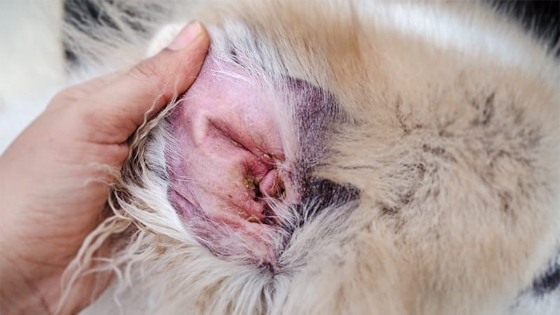
Malassezia dermatitis and/or otitis are common in dogs and the problem is overrepresented in the West Highland White Terrier, Basset Hound, Dachshund, and Cocker Spaniel. Pruritus and erythematous skin are the predominant clinical signs. With chronicity of pruritic behaviors, the skin becomes lichenified, excoriated, hyperpigmented and alopecic. Affected body sites may include the ear canals, lip folds, rostral chin, facial folds, axillae, inguinal skin, perineum, interdigital skin, and claw folds. Dogs may have a dry or greasy hair coat and malodor.
In cats, Malassezia dermatitis is rare. It is thought to be associated with severe underlying diseases such as endocrinopathies, metabolic diseases, neoplasias, feline leukemia virus, feline immunodeficiency virus, and hypereosinophilc syndrome. Certain breeds have an overgrowth unassociated with primary diseases (Devon Rex). In cats, clinical signs may include otitis, chin acne (a keratinization defect), paronychia (claw fold overgrowth), and erythematous greasy seborrhea. Pruritus may or may not be present.
How is Malassezia Dermatitis Diagnosed?
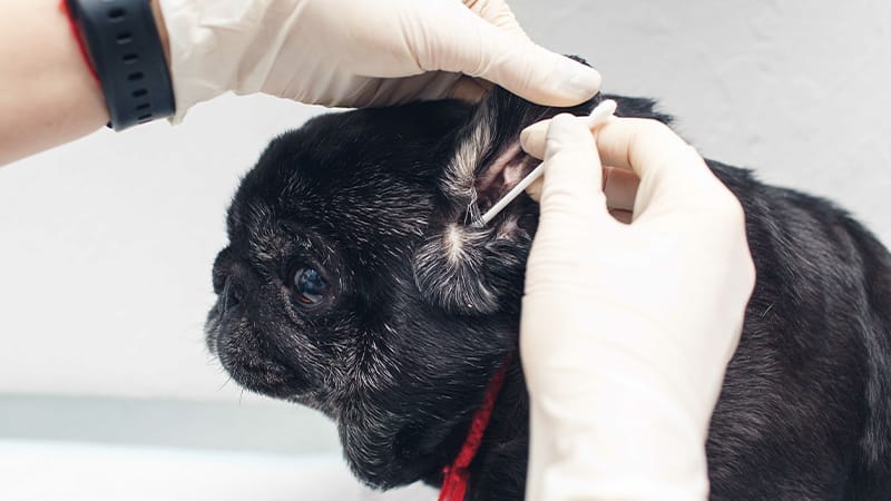
The diagnosis of Malassezia is relatively easy in practice by use of simple diagnostic tests. For quantitative evaluation, samples are collected for cytologic evaluation and stained. Samples may be collected from the ear canal or from affected skin sites. Typical sites include the claw fold, facial folds, lip folds, erythematous or lesional skin surfaces, and the ear canals. Salivary staining of the hair coat is common, and the underlying skin should be examined thoroughly for suggestive lesions.
Several techniques may be used for sample collection and include dry skin scrapes, impression smears, adhesive tape preparations, and use of cotton swabs. Specimens are placed upon a glass slide, stained and examined with a microscope. MP are peanut shaped budding yeasts that usually stain dark blue-purple. Interpretation of cytology requires consideration of other factors. Quantification of yeast number, assessment of clinical signs and historical features guide therapy choices. Normal animals and some body sites may have increased yeast numbers without any clinical signs. Furthermore, dogs allergic to the yeast may have significant symptoms even with very few yeast on the skin.
How is Yeast Dermatitis Treated?
Treatment recommendations usually involve a combination of systemic and topical therapy for generalized infections. Once the infection is controlled, topical therapy is modified and continued to decrease the likelihood of recurrence whilst other problems are addressed. One can also modify systemic therapy for long-term control (see below).
Topical therapy:
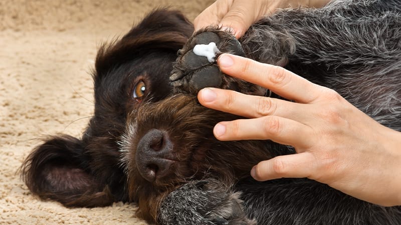
Topical therapy should be prescribed in all cases of yeast dermatitis for the purposes of mechanical removal of colonized keratinocytes from the skin surfaces. Shampoos are the most commonly used topical therapy. Active ingredients in shampoos with efficacy against MP include miconazole, ketoconazole, chlorhexidine at high concentration, and selenium sulfide (not in cats). Shampoos do not offer residual activity. Residual activity may be achieved with creams, lotions, and other leave-on products. Treatment of localized body sites such as skin folds may be managed with daily application of products containing miconazole, ketoconazole, fluconazole, chlotrimazole in the form of lotions, crèmes, wipes, and towelettes. Acetic acid (2.5%) is an inexpensive alternative, and is formulated by mixing 1 part water to 1 part vinegar.
Systemic therapy:
I consider use of systemic antifungal agents based upon patient characteristics and severity of infection. All cases with severe or generalized Malassezia dermatitis should be treated with oral medications. The duration of therapy using a systemic antifungal depends upon reevaluation of cytology of previously affected skin and improvement of clinical signs. The most common systemic medications for yeast include ketoconazole, itraconazole, fluconazole and terbinafine. Ketoconazole is extensively metabolized in the liver; the use in patients with hepatic disease is contraindicated. The most commonly observed side effects include vomiting, anorexia, and diarrhea. Ketoconazole should not be used in cats, as it is hepatotoxic. Evaluation of a serum biochemistry test is recommended prior to treatment with ketoconazole in most cases and every 1-3 months thereafter if ketoconazole is used for extended periods. Pulse administration of ketoconazole may also be used, and I use pulse schedule for maintenance whilst pursuing primary diseases or for control in cases where recurrence is common. Itraconazole is an antifungal agent also metabolized extensively in the liver. However, side effects are less frequent. This antifungal is widely used in cats. Dosage schedules vary and may involve once daily dosing or a pulsed schedule. Fluconazole is an antifungal agent that may be used in dogs and cats. Fluconazole is excreted in the kidney. Thus, fewer systemic gastrointestinal side effects are observed. Dosage schedules vary and may involve once daily dosing or a pulsed schedule. Terbinafine has been investigated for the treatment of yeast dermatitis. Terbinafine is an antifungal agent with low toxicity.